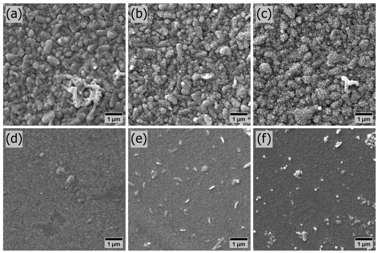
Figure 2.
SEM characterization of the surface morphology of electrodeposited and spin-coated PANI thin films. The micrographs were enlarged 10,000 times. The electrodeposited samples are presented on the top row and the spin-coated ones on the bottom row. J5T300, J5T600, and J5T1200 in (a–c), respectively. SC2000, SC1000, and SC500 in (d–f), respectively.

