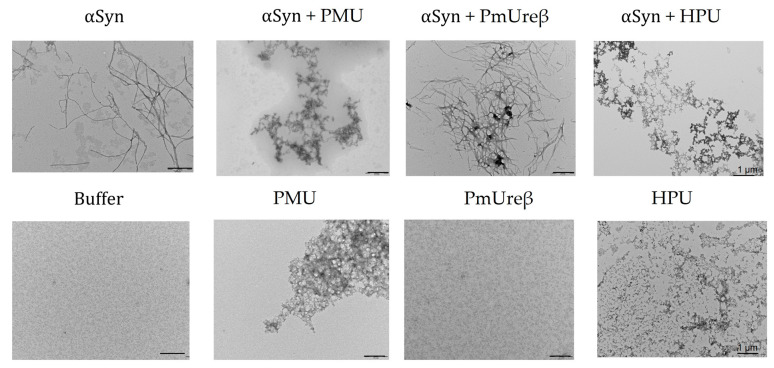
Figure 9.
TEM images of α-synuclein aggregates formed in the presence of ureases. The reaction mixtures contained 50 µM α-synuclein in the absence (bottom panels) or the presence (top panels) of 5 µM ureases (PMU, PmUreβ, or HPU) in 10 mM NaPB, pH 7.5, 100 mM NaCl (buffer). Incubation proceeded for 150 h under gentle agitation at 37 °C. Fibril formation was confirmed via transmission electron microscopy of the reaction mixtures. The microscopies are typical results of at least three different samples. Scales bar represents 500 nm, with the exception of αSyn + HPU and HPU (1 μm).

