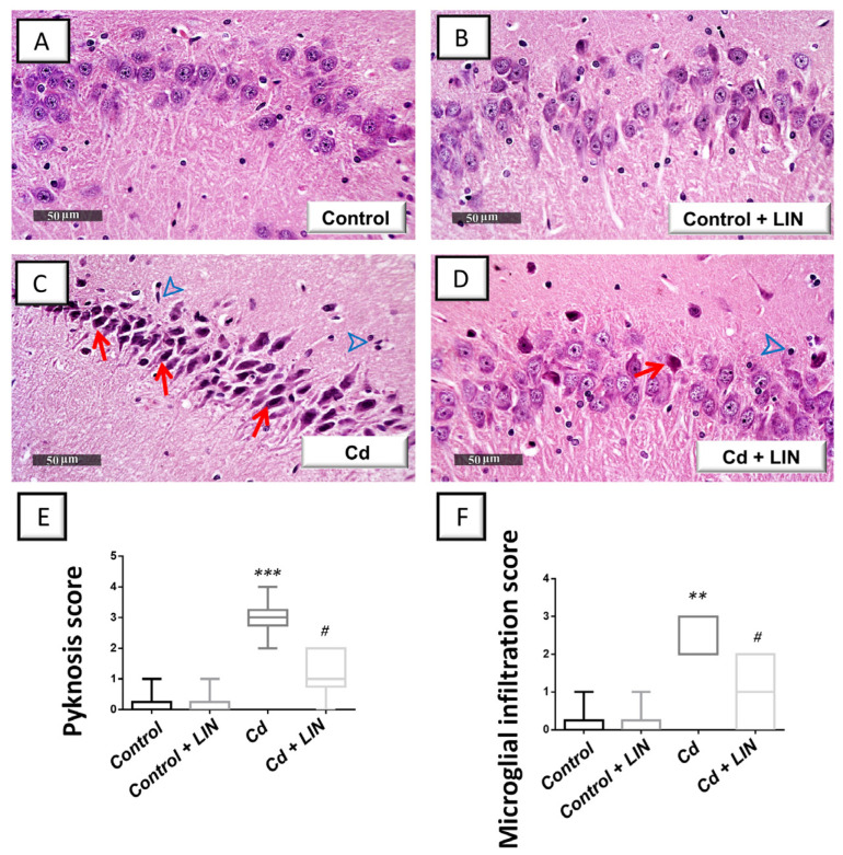
Figure 2.
Linagliptin mitigates cadmium-induced neuronal degeneration and glial cell infiltration in the hippocampi of rats. Hippocampal sagittal sections were inspected by a light microscope after staining with hematoxylin and eosin (H-E). An intact structure of the hippocampus and organized distribution of the hippocampal layers were seen in the control (A) and control + linagliptin groups (B). Moreover, the hippocampus area of both groups revealed intact pyramidal neurons with distinct nuclear and subcellular details. (C) The hippocampus area of Cd group showed marked degeneration, neuronal loss, pyknotic pyramidal neurons with invisible subcellular details (red arrow), and reactive microglial cell infiltrates (arrowhead). (D) Cd + linagliptin group demonstrated attenuation of the hippocampal pathological changes and demonstrated few records of neuronal degenerative changes (red arrow). However, some reactive glial cell infiltrates were still seen (arrowhead). (E,F) The scores of pyknosis and microglial cell infiltration were significantly lowered by linagliptin administration to cadmium-intoxicated animals. N = 6 in each group (median with interquartile range). A p-value of less than 0.05 was significant. ** p < 0.01, or *** p < 0.001, compared to control; # p < 0.05, compared to cadmium (Dunn’s test for multi-comparisons and Kruskal–Wallis test). Cd, cadmium chloride; LIN, linagliptin.

