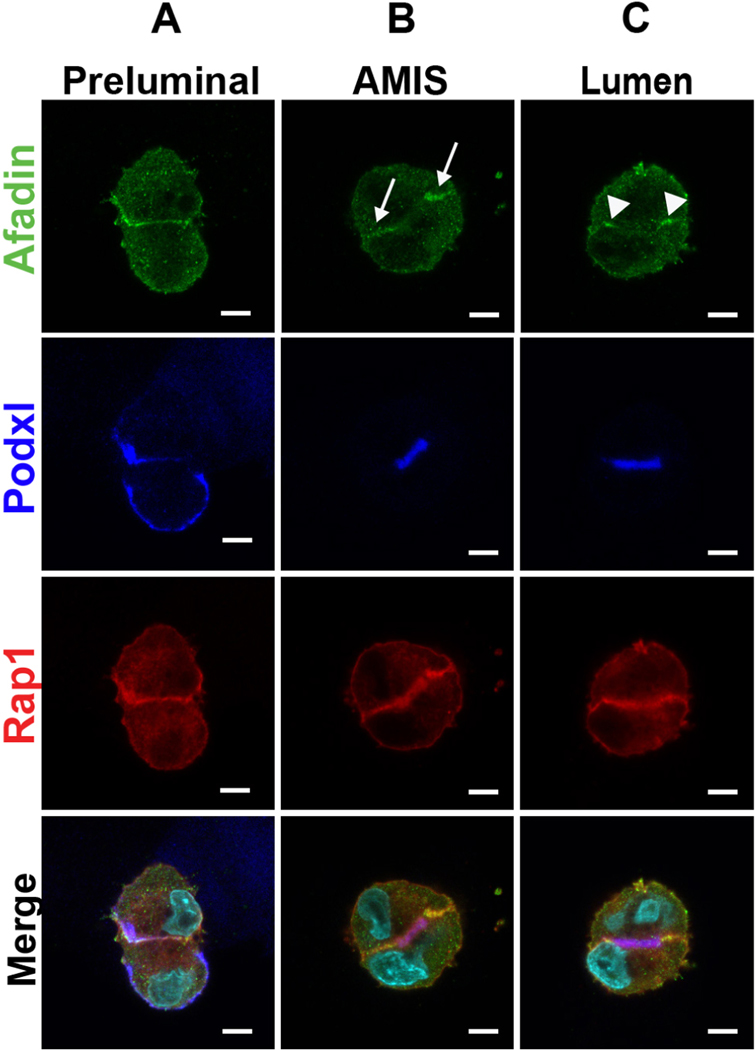
Figure 1. mCherry-Rap1a and Afadin localization during stages of lumen formation.
A. Immunofluorescence of two-cell stage MDCK spheroids with Afadin (green), mCherry-Rap1a (red), and podxl (blue) prior to lumen formation.
B. Immunofluorescence as in A at the apical membrane initiation site (AMIS) stage.
C. Immunofluorescence at the lumen stage.
Arrows depict Afadin at lateral membranes and arrowheads show Afadin at apical-lateral junctions. Results are representative of at least 2 independent experiments. Scale bars: 5 μm.

