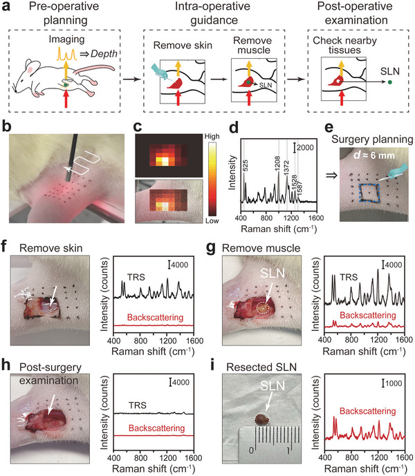
Figure 6.

Intraoperative detection and minimal surgery of the rat SLN using SETRS. a) The schematic illustration of the perioperative surgery guidance process. b–e) Pre‐operative planning, with b) non‐invasive TRS imaging process (n = 3), c) corresponding TRS image and its overlay with the bright‐field image. d) Raman spectrum at the brightest spot of the TRS image. e) Pre‐operative surgical planning based on TRS imaging and depth prediction results. f–i) The TRS and backscattering Raman measurements (n = 3): f) after the removal of skin and subcutaneous fat, g) after the removal of muscle to expose the lymph node in situ, h) the remaining tissue after the lymph node is resected, and i) the resected lymph node. The TRS and backscattering Raman measurements were conducted with the laser irradiances of 0.105 and 1.31 J cm−2, and the integration time of 0.5 s and 10 ms, respectively.
