
Figure 2.
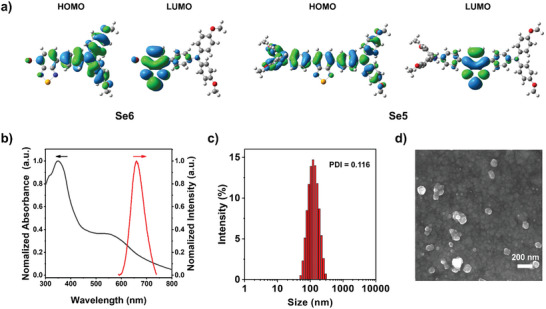
a) The DFT calculated electron cloud distributions in HOMOs and LUMOs of Se6 and Se5. b) Normalized absorption and fluorescence spectra of Se6‐NPs in aqueous solution (10 µm). c) The particle size distribution of Se6‐NPs detected from dynamic light scattering (DLS). d) Scanning electron microscopy (SEM) images of Se6‐NPs, scale bar: 200 nm. The polydispersity index (PDI) of nanoparticles was also inset in (c).