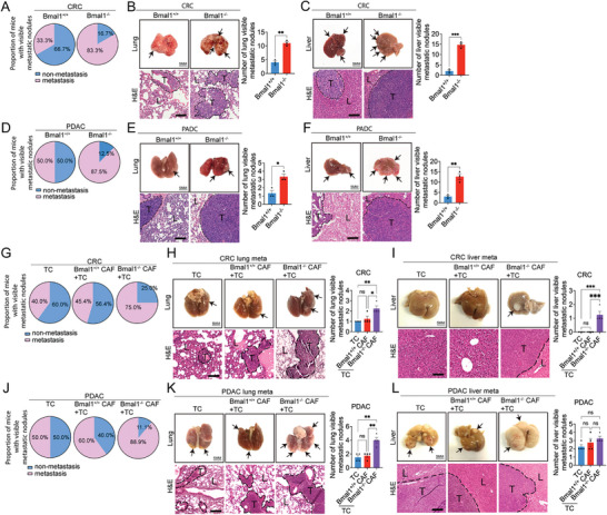
Figure 6.

Genetic deletion of Bmal1 promotes cancer metastasis. A) Quantification of visible metastases on the surface of lungs and livers in CRC tumor‐bearing Bmal1+/+ and Bmal1−/− mice (n = 6 mice per group). B,C) Gross examination and H&E staining of lungs and livers of CRC tumor‐bearing Bmal1+/+ and Bmal1−/− mice (scale bar = 5 mm). Quantification of visible metastatic nodules by H&E staining (n = 3 random fields per group). D) Quantification of visible metastases on the surface of lungs and livers in PDAC tumor‐bearing Bmal1+/+ and Bmal1−/− mice (n = 8 mice per group). E,F) Gross examination and H&E staining of lungs and livers of PDAC tumor‐bearing Bmal1+/+ and Bmal1−/− mice (scale bar = 5 mm). Quantification of visible metastatic nodules by H&E staining (n = 3 random fields per group). G) Quantification of visible metastases on the surface of lungs and livers in C57BL/6 wild type mice that received CRC tumor cells, CRC tumor cells plus Bmal1+/+ CAFs, and CRC tumor cells plus Bmal1−/− CAFs implantation (n = 10 mice per group). H,I) Gross examination and H&E staining of lungs and livers of CRC tumor‐bearing in C57BL/6 wild type mice that received CRC tumor cells, CRC tumor cells plus Bmal1+/+ CAFs, and CRC tumor cells plus Bmal1−/− CAFs implantation (scale bar = 5 mm). Quantification of visible metastatic nodules by H&E staining (n = 4 random fields per group). J) Quantification of visible metastases on the surface of lungs and livers in PDAC tumor‐bearing in C57BL/6 wild type mice that received PDAC tumor cells, PDAC tumor cells plus Bmal1+/+ CAFs, and PDAC tumors cell plus Bmal1−/− CAFs implantation (n = 10 mice per group). K,L) Gross examination and H&E staining of lungs and livers of PDAC tumor‐bearing in C57BL/6 wild type mice that received PDAC tumor cells, PDAC tumor cells plus Bmal1+/+ CAFs, and PDAC tumor cells plus Bmal1−/− CAFs implantation (scale bar = 5 mm). Quantification of visible metastatic nodules by H&E staining (n = 4 random fields per group). Data presented as mean ± S.E.M. *P < 0.05; **P < 0.01; ***P < 0.001, ns = not significant; two‐tailed student t‐test and one‐way ANOVA. TC = tumor cell. Apparent metastatic nodules are indicated by arrows. Dashed lines encircle cancer metastases. T = tumor; L = lung or liver. H&E scale bar = 100 µm.
