FIGURE 2.

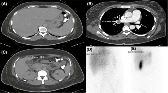
CT scan of (A) the abdomen depicting splenomegaly (B) of the chest showing an enlarged right hilar lymph node and (C) the abdomen showing a thickened gallbladder wall. (D and E) are HIDA scans showing the absence of acute cholecystitis.

CT scan of (A) the abdomen depicting splenomegaly (B) of the chest showing an enlarged right hilar lymph node and (C) the abdomen showing a thickened gallbladder wall. (D and E) are HIDA scans showing the absence of acute cholecystitis.