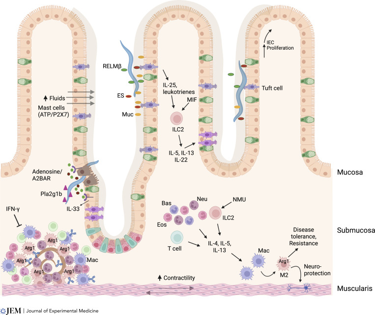
Figure 3.
Multiple mechanisms drive host protection to helminths in intestinal tissue. The type 2 response is likely initiated by tuft cell sensing of helminth excretory/secretory (ES) products in the lumen. This triggers tuft cell production of leukotrienes, which, combined with constitutive tuft cell IL-25 production, and also production of macrophage migration inhibitory factor (MIF) by unknown sources and neuromedin U (NMU), induces type 2 responses. Helminths that actually invade the epithelial barrier damage intestinal epithelial cells (IECs), which release ATP. Extracellular ATP, when degraded to ADP, can bind A2BAR, which activate IECs contributing to tuft cell hyperplasia and IL-33 production. ATP may also bind P2X7 on mast cells. Myeloid cells and ILC2s produce type 2 cytokines driving the response. As parasites cross the intestinal barrier, group 1b phospholipase A2 (Pla2g1b) binds the parasites, impairing their development. H. polygyrus larvae dwell in the submucosa as they develop into adults. After secondary inoculation, a rapidly developing granuloma, composed primarily of M2 macrophages, and also eosinophils, surrounds the parasite and impairs its development through Arg1-dependent mechanisms. Type 2 cytokines drive the type 2 granulomas while IFN-γ suppresses it. Helminths in the lumen may be expulsed through a combination of fluids flowing into the lumen, enhanced by activated mast cells, production of mucins (muc), and increased muscle contractility (weep-and-sweep response). Also, parasites, such as Trichuris, residing in the epithelial layer may be expulsed through upward IEC proliferation. M2 macrophages also mediate enteric disease tolerance mechanisms including neuronal protection.

