
Figure 2.
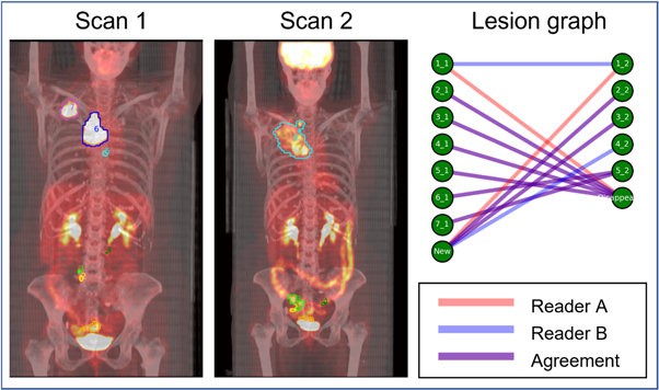
Lesion matching in a female subject with stage III NSCLC imaged with 18F-FDG PET/CT before (Scan 1) and after (Scan 2) platinum-based chemotherapy. PET/CT were acquired 179 d apart. The lesion located in the rectum (orange contour, label 1) is matched differently between readers. Reader A determines that the lesion disappears after scan 1 and a new lesion in a similar area appears on scan 2. Reader B determines that these two lesions are homologous and should be matched between scans.