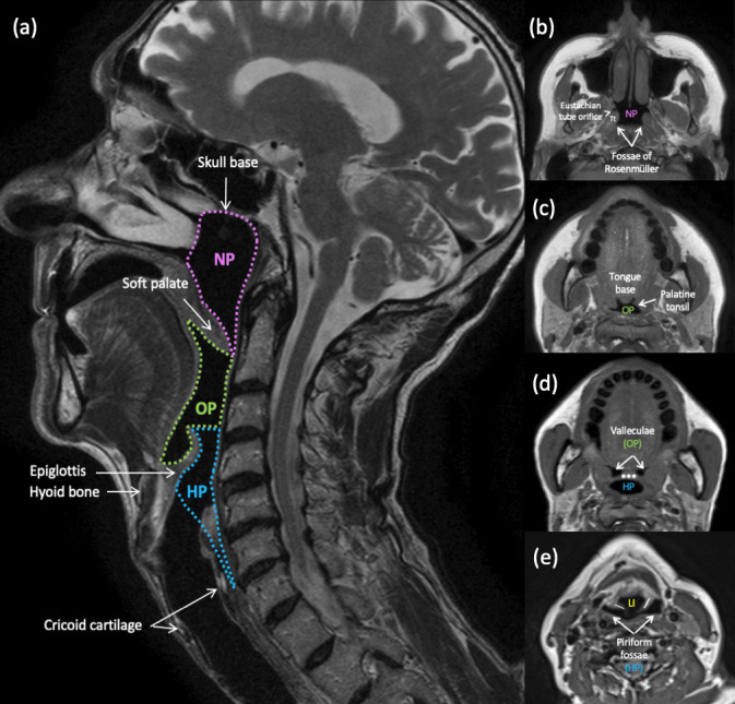
Figure 1.

Pharyngeal anatomy. (a) Anatomical divisions of the pharynx and their boundaries. Sagittal T2-weighted MRI. Nasopharynx (NP): skull base to the free edge of the soft palate. Oropharynx (OP): free edge of the soft palate to the hyoid bone. Hypopharynx (HP): hyoid bone to the inferior border of the cricoid cartilage or cricopharyngeus muscle.(b-e) Important anatomical landmarks within each subdivison. Axial T1-weighted MR images through the pharynx. (b) Nasopharynx; Tt = torus tubarius; (c, d) Oropharynx; *** = epiglottis; (d,e) Hypopharynx; \ / = aryepiglottic folds; LI = laryngeal inlet. Note the complex anatomy at the oropharyngeal-hypopharyngeal-supraglottic junction, where from one cavity (OP), two separate cavities arise (HP and larynx).
