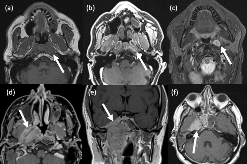
Figure 10.

Nasopharyngeal carcinoma. (a-c) Small NPC in the left fossa of Rosenmüller. A 66-year old male presented with a suspicious left nasopharyngeal lesion on nasendoscopy. (a) Axial T1-weighted STIR MRI shows asymmetrical enlargement of the left nasopharynx. (b) Axial T1-weighted post-contrast MRI shows heterogeneous enhancement. (c) Axial T2-weighted, fat-suppressed MRI shows an enlarged ipsilateral retropharyngeal node – a common region of nodal metastasis in NPC. (d-f) Invasive NPC. A 48-year old male presented with a large, highly suspicious right nasopharyngeal mass on nasendoscopy. (d) Axial fat-suppressed T1-weighted, post-contrast MRI shows an irregular, heterogeneously enhancing, right nasopharyngeal mass and middle ear effusion. (e) Coronal T1-weighted, post-contrast MRI shows the craniocaudal extent of the mass, with skull base invasion and intracranial extension. (f) Axial T1-weighted, post-contrast MRI Brain shows cranial invasion into the right middle cranial fossa.
