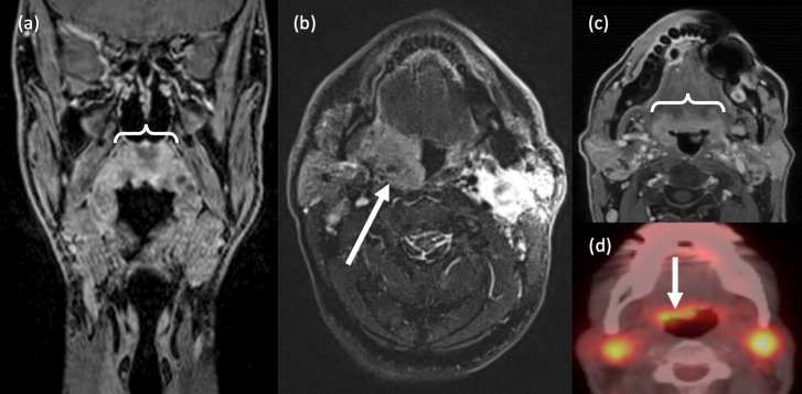
Figure 11.
Oropharyngeal SCC. (a) Soft palate SCC. Coronal T1-weighted, post-contrast MRI shows a large, ill-defined, enhancing soft palate mass which extends laterally to the tonsillar pillars, more notably on the right. (b) Tonsillar SCC. Axial T2-weighted, fat-suppressed MRI shows a large right palatine tonsil mass. Histology confirmed a poorly undifferentiated SCC. (c,d) Base of tongue SCC. A 64-year old male who presented with cervical lymphadenopathy underwent a lymph node biopsy which confirmed metastatic SCC. (c) Axial T1-weighted, post-contrast MRI shows mild asymmetrical thickening at the lingual tonsils and right base of tongue. (d) Axial FDG-PET CT confirms abnormal uptake at the base of tongue on the right, as well as in bilateral cervical lymph nodes.

