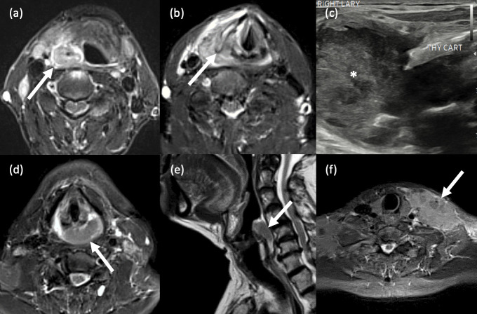
Figure 12.
Hypopharyngeal SCC. (a-c) Pyriform sinus SCC. Axial STIR MR images show (a) a large right hypopharyngeal mass lesion which is centred around the right piriform fossa, extends anterolaterally into the surrounding superficial soft tissues and; (b) anteromedially and caudally into the ipsilateral aryepiglottic fold and supraglottic larynx. The right thyroid cartilage is partially destroyed. (c) B-mode ultrasound imaging in the same patient shows a large, right-sided heterogeneous mass (*) abutting and protruding outwards beyond the destroyed right thyroid cartilage (labelled). (d-f) Posterior pharyngeal wall SCC. A 52-year old female presented with a 3-month history of dysphagia and weight loss. (d) Axial STIR MRI shows a large lesion in the mid and left posterior wall of the hypopharynx. (b) Sagittal T2-weighted MRI better demonstrates that the posterior wall is the subsite around which the mass is centred, in addition to its craniocaudal extent. (f) Axial STIR MRI in the same patient shows a large, irregular left lower cervical nodal mass (extranodal spread).

