Figure 13.

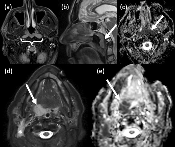
Pharyngeal lymphomas. (a-c) Nasopharyngeal lymphoma. A 69-year old male presented with a post-nasal space mass and bilateral cervical lymphadenopathy. (a) Axial T2-weighted, fat suppressed MRI reveals a bulky, symmetrical, slightly hyperintense mass in the roof of the nasopharynx. Extensive bilateral cervical lymphadenopathy was confirmed (not shown). (b) Sagittal T2-weighted, fat-suppressed MRI shows the craniocaudal extent of the mass, i.e. the entire length of the posterior nasopharynx. There is no evidence of skull base invasion. (c) Axial EPI DWI and relative ADC map show marked restricted diffusion and low ADC values. Histopathology confirmed diffuse large B cell lymphoma (DLBCL). (d,e) Oropharyngeal lymphoma. (d) Axial T2-weighted, fat-suppressed MRI reveals a relatively homogeneous right tonsillar lesion and an abnormal right level 2 lymph node (*). (e) Axial EPI DWI ADC imaging in the same patient shows well the restricted diffusion within the lesion.
