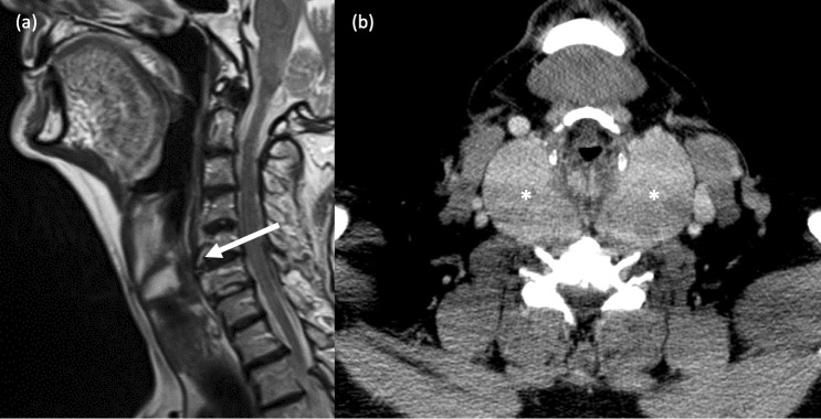
Figure 15.
Extrinsic compression of the hypopharynx. (a) Cervical vertebral osteophytosis. An elderly patient presented with mild dysphagia. Sagittal T2-weighted MRI of the neck shows severe anterior osteophytosis of the C4-6 vertebral bodies resulting in indentation of the posterior hypopharyngeal wall. (b) Multinodular goitre. A 60-year old male with dysphagia and clinically obvious multinodular goitre required cross-sectional imaging prior to surgical intervention. Axial, contrast-enhanced CT Neck image shows a large, bilateral multinodular thyroid goitre (* *) causing marked compression of the hypopharynx.

