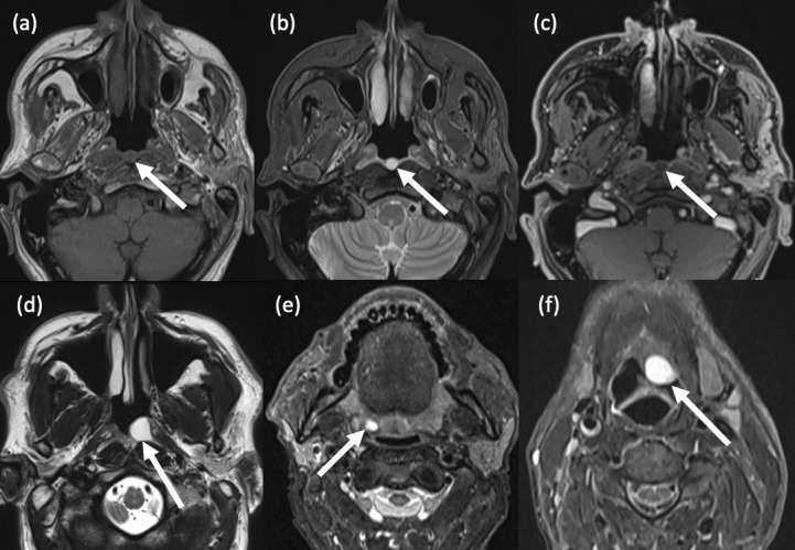
Figure 2.

Pharyngeal cysts. (a-c) Tornwaldt cyst. (a) Axial T1-weighted TSE MRI shows a well-circumscribed, round, hypointense lesion in the posterior nasopharyngeal midline; (b) Axial T2-weighted TSE MRI shows homogeneous central hyperintensity; (c) Axial T1-weighted, fat suppressed, post-contrast MRI shows no enhancement within the lesion.(d) Nasopharyngeal mucous retention cyst. Axial T2-weighted TSE MRI shows a hyperintense cyst in the left fossa of Rosenmüller. (e) Tonsillar cyst. Axial STIR MRI shows an incidental small right palatine tonsillar retention cyst. (f) Vallecular cyst. A 60-year-old male presented with globus sensation and throat irritation for three weeks. Axial STIR MRI shows a large, well-defined cyst which expands the left vallecula and abuts the epiglottis.
