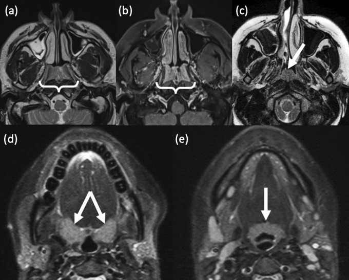
Figure 3.

Benign lymphoid hyperplasia. (a,b) Nasopharyngeal lymphoid hyperplasia in a 41-year-old male smoker. (a) Axial T2-weighted MRI shows symmetrical mucosal thickening in the roof of the nasopharynx. (b) Axial T1-weighted, post-contrast MRI demonstrates a vertical stripe-like enhancing pattern (bright and dark). Tiny, non-enhancing cysts are also noted within the lymphoid tissue. (c) Focal adenoid tonsillar hyperplasia in an adolescent as demonstrated on axial T2-weighted MRI. (d,e) Oropharyngeal lymphoid hyperplasia. A 32-year old female presented with persistently enlarged, bilateral upper cervical lymph nodes. Axial T2-weighted STIR MR images show diffuse, symmetrical enlargement of both the (d) palatine and (e) lingual tonsils.
