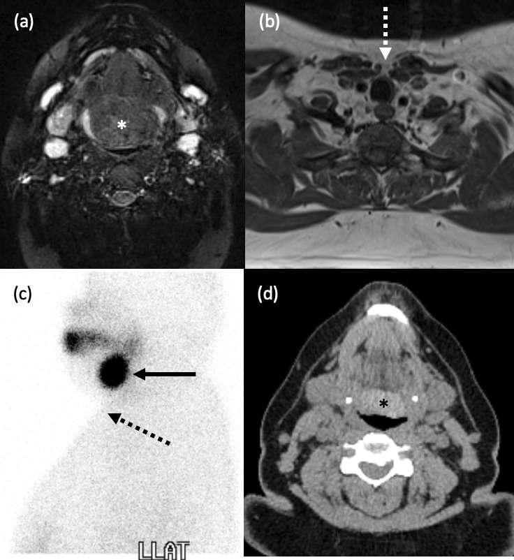
Figure 5.

Lingual thyroid. (a-d) A 24-year old female presented with a large tongue base mass. (a) Axial T2-weighted, fat suppressed MRI demonstrates a lobulated, heterogeneous lesion at the base of the tongue (*). (b) Axial T1-weighted MRI confirmed the absence of any normal thyroid tissue in the pretracheal neck (white dashed arrow). (c) Left lateral view from a Tc-99m pertechnetate thyroid scintigraphy study shows intense uptake at the tongue base (black solid arrow), and none in the usual position (black dashed arrow). (d) Axial, unenhanced CT Neck over 10 years later in the same patient shows a hyperdense mass typical of thyroid tissue at the tongue base (*).
