Figure 9.

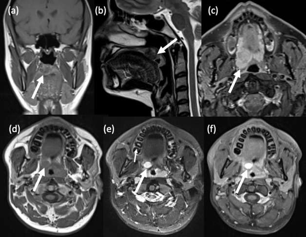
Benign pharyngeal tumours. (a,b) Minor salivary gland pleomorphic adenoma. A 36-year old female presented with a lump in her soft palate. (a) Coronal T1-weighted, pre-contrast MRI shows a well-circumscribed, heterogeneous lesion within the right soft palate which is predominantly hypointense. Smaller lesions are often homogeneously hypointense. (b) Sagittal T2-weighted MRI shows a hypointense rim surrounding the lesion, which represents the fibrous capsule that is characteristic of pleomorphic adenomas. Histology confirmed pleomorphic adenoma with extensive epidermoid differentiation. (c) Minor salivary gland pleomorphic adenoma. A 61-year old male also presented with a soft palate swelling. Axial T1-weighted, fat-suppressed, post-contrast MRI reveals a well-defined, heterogeneously enhancing lesion in the soft palate. Another common feature of these tumours, which is also seen here, is its lobulated contour. (d-f) Schwannoma. A 17-year old male presented with a 1cm lesion in his right soft palate. (d) Axial T1-weighted, pre-contrast MRI shows a well-defined, ovoid lesion in the right soft palate which is isointense to muscle. Note also the thin, peripheral rind of fat (the so-called ‘split fat’ sign). (e) Axial T2-weighted, fat-suppressed MRI shows intralesional hyperintensity. (f) Axial T1-weighted, fat-suppressed, post-contrast MRI shows intense contrast enhancement, a typical imaging feature of schwannoma. Though not demonstrated in this case, the ‘fascicular’ sign may also be seen, i.e. multiple ring-like structures within the lesion which reflect fascicular bundles.
