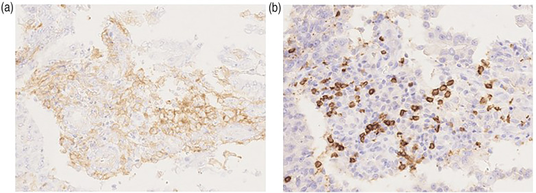
Figure 6.
Representative immunohistochemistry images for the PD-L1 and CD8 cells. The epithelial compartment is positive for (a) PD-L1, whereas the infiltrating immune cells are positive for (b) CD8. The images are shown at 200× magnification.
PD-L1: programmed cell death ligand 1; CD8: cluster of differentiation 8.

