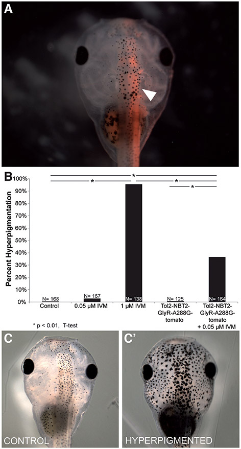
Fig. 2. Driving GlyR-A288G expression in neural tissue is sufficient to induce hyperpigmentation.
Embryos were injected with Tol2-NBT2-GlyR-A288G-tomato (driving the neural-specific expression of a GlyCl channel with increased sensitivity to ivermectin fused to a tdTomato fluorescent reporter) into 1 cell of 2-cell embryo (NF stage 2). (A) Neural-specific expression was confirmed in NF stage 45 tadpoles (white arrow). (B) Embryos that had been injected with Tol2-NBT2-GlyR-A388G-tom and treated with 0.05 μM ivermectin displayed significant levels (36.3%, N=164, P<<0.01 in injected & treated embryos) of hyperpigmentation (C’) compared to control embryos (C) and those that had been injected and not exposed to ivermectin (0.8% hyperpigmented, N=125).

