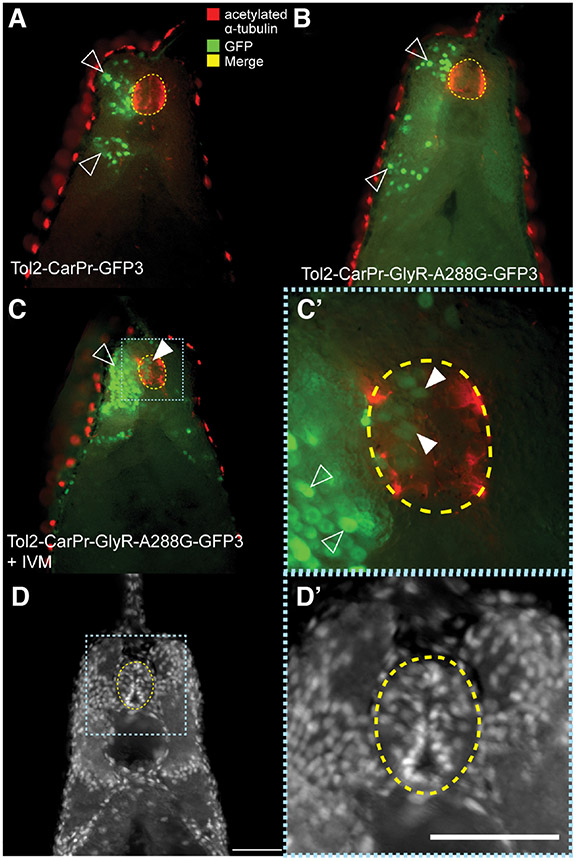
Fig. 5. Mis-localized Car promoter-driven GFP+ cells in neural tube are not completely neural.
(A) Double labeling co-immunohistochemistry was performed on sections of tailbud stage (NF stage 28/29) Xenopus embryos that had been injected with Tol2-CarPr-GFP3 using an anti-GFP antibody to track cells that have been activated by the cardiac actin promoter alongside an anti-acetylated α-tubulin, labeling neural tissue. This revealed normal expression of GFP in the somatic tissue. (B) Co-immunos on sections of embryos that had been injected with a Tol2-CarPR construct driving expression of hypersensitive GlyR-A288G mutant also revealed normal localization of injected construct. (C) Embryos injected with Tol2-CarPr-GlyR-A288G-GFP3 and treated with 0.05 μM ivermectin displayed abnormal neural localization (white arrow), however, the GFP+ cells in the neural tube did not co-localize with acetylated α-tubulin cells; white arrows in (C’). (D,D’) Hoechst nuclear staining of sections.

