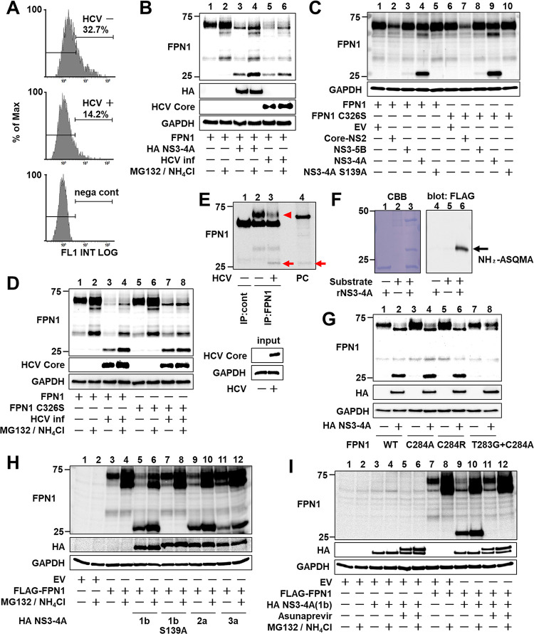
Fig 4. Proteolytic cleavage of FPN1 mediated by HCV NS3-4A protease.
(A) Huh7.5.1 cells with or without HCV infection 1 day after transfection of the FPN1-expression plasmid were harvested at 1 dpi and reacted with anti-FPN1 antibody. The percentage of FPN1-positive cells was counted by flow cytometry. Cells transfected with an EV (nega cont) were used as a negative control. The figure is representative of three experiments with similar results. (B, D) At 1 dpi, cells with or without HCV infection were transfected (B) with the FPN1-expression plasmid together with the NS3-4A expression plasmid or the empty vector, (D) with plasmids expressing FPN1 or FPN1 C326S. At 2 dpt, FPN1, HCV Core, and GAPDH were analyzed by western blotting. (C) FPN1 and GAPDH were analyzed by western blotting with cell lysates transfected with both plasmids expressing FPN1 or FPN1 C326S and Core-NS2, NS3-5B, NS3-4A, or NS3-4A S139A at 2 dpt. (E) HCV-infected cells 3 dpi were immunoprecipitated with anti-FPN1 antibody (BMP033) or rabbit IgG, followed by western blotting using anti-FPN1 antibody (NBP1-21502). HCV Core and GAPDH in input samples were assessed by western blotting without immunoprecipitation. A cell lysate transfected with plasmids expressing FPN1 and NS3-4A was used as a positive control (PC). Endogenous full-length- and processed FPN1 are indicated as an arrowhead and arrow, respectively. (F) MBP-FPN1 (aa 278–292)-sfGFP-FLAG fusion protein as a substrate was reacted with recombinant NS3-4A. After SDS-PAGE and transfer to a PVDF membrane, the processed band stained with CBB (left) was excised, followed by N-terminal analysis. Processing of the substrate fusion protein was confirmed by western blotting using anti-FLAG antibody (right). (G-I) FPN1, HA-tagged NS3-4A, and GAPDH were analyzed by western blotting. Cells were transfected with (F) the plasmid expressing WT- or mutated FPN1 together with the expression plasmid for HA-tagged NS3-4A or EV, (H) the FLAG-FPN1-expressing plasmid together with the HA-NS3-4A expressing plasmid from genotypes 1b, 2a, or 3a, and (I) the plasmids expressing FLAG-FPN1 and HA-NS3-4A in the presence or absence of 1 μM asunaprevir. (B, D, H, I) MG132 and NH4Cl were added at final concentrations of 10 μM and 10 mM, respectively, 12 h before cell harvesting.

