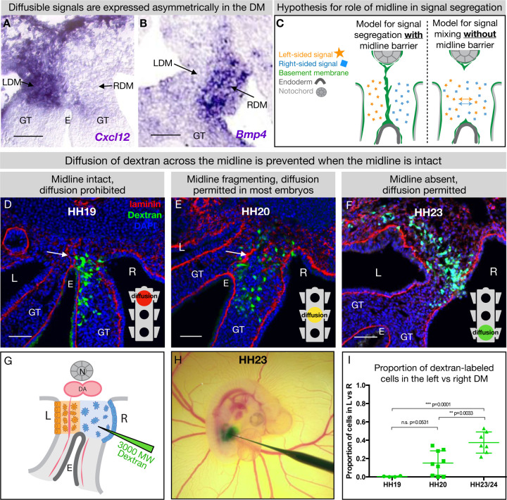
Figure 6. The DM midline serves as a barrier against diffusion.
(A, B) Genes encoding diffusible signals including Cxcl12 and Bmp4 are expressed asymmetrically in the DM. (C) Hypothesis for the role of the midline in limiting diffusion of left and right signals across DM. (D) At HH19, the midline is intact (white arrow) and diffusion of 3000 MW dextran (green) is limited to the right side. n = 4/4. (E) At HH20, the midline (white arrow) has begun to fragment. Diffusion across the midline is prohibited in some embryos (n = 2/9) but permitted in others (n = 7/9). (F) At later stages when the midline has disappeared, diffusion is allowed through the DM (n = 7/7). (G) Schematic of dextran injections into the right DM. (H) A dextran injection being performed on an HH23 embryo in ovo. (I) Proportion of dextran-labeled cells in the left vs. right DM, with unpaired t test. Scale bars = 50 um. LDM = left dorsal mesentery. RDM = right dorsal mesentery. GT = gut tube. E = endoderm. L = left. R = right. N = notochord. DA = dorsal aorta.

