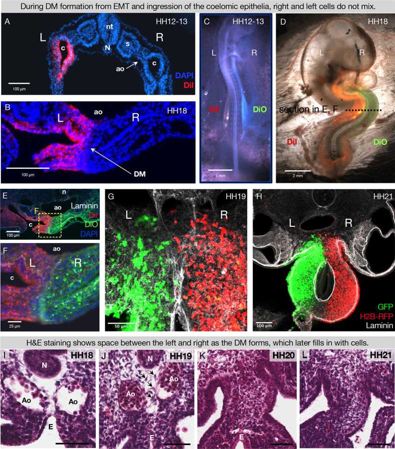
Figure 2. During DM formation from EMT and ingression of the coelomic epithelia, right and left cells do not mix.
When the coelomic cavity is injected with DiI at HH12–13, n = 5 (A), the labeled cells give rise to the mesenchymal and epithelial cells of the DM on the corresponding side of the embryo, n = 5 (B). When DiI and DiO are injected at HH12–13 into left and right coeloms, respectively, n = 6 (C), labeled cells are still segregated at HH18, n = 6 (D, E and F). The same results are found when cells are labeled by electroporation with pCAG-GFP (left) and pCl-H2B-RFP (right) (G and H), both when the midline is continuous (HH19 n = 3, G) and once it has disappeared (HH21 n = 3, H). (I-L) H&E staining of the DM at HH18 n = 5 (I) shows “empty space” between the notochord, endoderm, and dorsal aortae. At HH19 n = 5 (J), this space gains some cells (arrows), and the space is completely filled in by HH20 n = 4 (K), and HH21 n = 3 (L). Scale bars = 60 μm. nt = neural tube, c = coelom, ao = aorta, N = notochord, s = somite, DM = dorsal mesentery, L = left, R = right.

