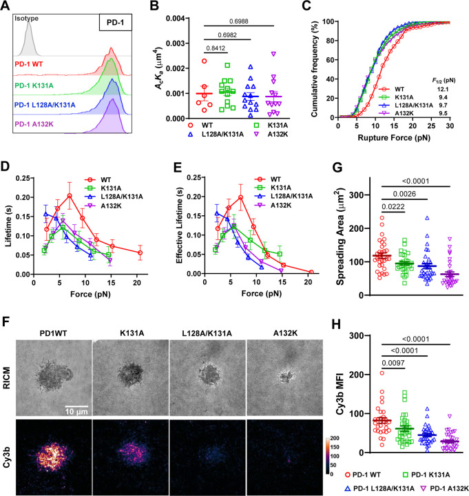
Fig. 5.
PD-1 mutants preventing force-induced atomic contacts impairs PD-1–PD-L2 mechanical stability. (A) Flow cytometry histograms comparing PD-1 staining of CHO cells expressing WT or indicated mutants of PD-1. (B) 2D effective affinity of PD-L2 binding to CHO cells expressing WT or indicated mutants of PD-1. n = 6, 12, 13, and 12 cell pairs for WT, K131A, L128A/K131A, and A132K, respectively. (C) Cumulative frequencies of rupture force events for PD-L2 binding to PD-1 WT (n = 278 events), K131A (n = 210 events), L128A/K131A (n = 345 events), and A132K (n = 270 events) bonds. p < 0.0001 comparing F1/2 of WT and each mutant using two-tailed Mann-Whitney test. (D) Mean ± sem bond lifetime vs force plots for single PD-L2 bonds with PD-1 WT (n = 785 events), K131A (n = 625 events), L128A/K131A (n = 759 events), and A132K (n = 780 events). p < 0.0001 comparing lifetime vs force distributions of WT and each mutant using two-tailed two-dimensional Kolmogorov-Smirnov test. (F) Representative RICM and Cy3b fluorescence images of CHO cells expressing PD-1 WT or indicated mutants 30 min after landing on glass surface functionalized with PD-L2-coupled MTP of 4.7 pN threshold force. (G-H) Quantification of cell spreading area (G) and tension signal (H) for conditions in F. n = 29, 30, 30, and 30 pooled from 3 independent experiments. Numbers on graphs represent p values calculated from two-tailed student t-test.

