
FIG. 2.
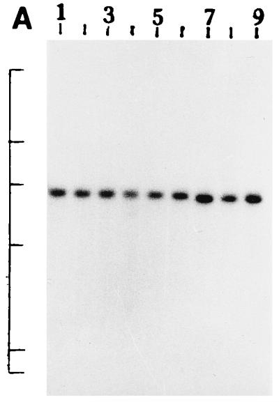
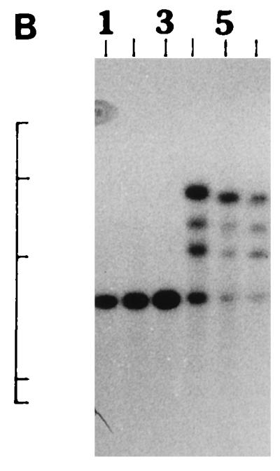
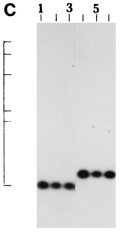
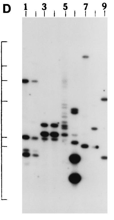

(A) Southern blot hybridization of PstI-digested non-O1, non-O139 V. cholerae chromosomal DNA with the hlyA probe. Lanes 1 to 9, strains AM107, AS61, AM109, AM111, AM112, AM113, AS64, AS67, and AS68, respectively. The positions of bacteriophage λ HindIII molecular size markers run on the same gel are indicated by bars from top to bottom (23.13, 9.41, 6.55, 4.36, 2.32, and 2.0 kb). The patterns for only representative strains are shown here. (B) Southern blot hybridization of XbaI-BglII-digested non-O1, non-O139 V. cholerae chromosomal DNA with the hlyU probe. Lanes 1 to 3, strains AS61, AM112, and AS64 (strains showing the HUI pattern), respectively; lanes 4 to 6, strains AM107, AM108, and AS66 (strains showing the HUII pattern), respectively. The positions of bacteriophage λ HindIII molecular size markers run on the same gel are indicated by bars from top to bottom (9.41, 6.55, 4.36, 2.32, and 2.0 kb). The patterns for only representative strains are shown here. (C) Southern blot hybridization of Sau3AI-digested non-O1, non-O139 V. cholerae chromosomal DNA with the hlx probe. Lanes 1 to 3, strains AM109, AS64, and AS68 (strains showing the HLI pattern), respectively; lanes 4 to 6, strains AM107, AS60, and AS61 (strains showing the HLII pattern), respectively. The positions of bacteriophage λ HindIII molecular size markers run on the same gel are indicated by bars from top to bottom (9.4, 6.55, 4.36, 2.32, 2.0 and 0.56 kb). The patterns for only a few representative strains are shown. (D) Southern blot hybridization of HindIII-digested non-O1, non-O139 V. cholerae chromosomal DNA with the attRS1 probe. Lanes 1 and 2, strains AM107 and AS61 (strains the showing A1 pattern), respectively; lanes 3 and 4, strains AS64 and AS68 (strains showing the A2 pattern), respectively; lane 5, strain AS66 (showing a partial digest); lanes 6 to 9, strains showing unique patterns (strains AM109, AM111, AM112, and AM113, respectively). The positions of bacteriophage λ HindIII molecular size markers run on the same gel are indicated by bars from top to bottom (23.13, 9.41, 6.55, 4.36, 2.32, 2.0, and 0.5 kb). The patterns for representative strains are shown. (E) Southern blot hybridization of PstI-digested non-O1, non-O139 V. cholerae chromosomal DNA with the toxR probe. Lanes 1 to 4, strains AM107, AM108, AS60, and AS61 (strains showing the TR1 pattern), respectively; lanes 5 to 8, strains AM109, AM113, AS64, and AS68 (strains showing the TR3 pattern), respectively; lanes 9 and 10, strains AM111 and AM112 (strains showing the TR2 pattern), respectively. The positions of bacteriophage λ HindIII molecular size markers run on the same gel are indicated by bars from top to bottom (9.41, 6.55, 4.36, 2.32, and 2.0 kb). The patterns for only representative strains are shown.