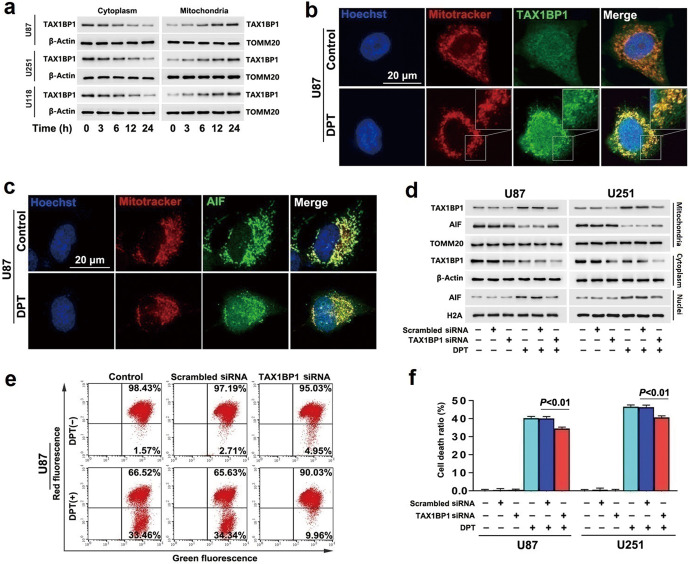
Fig. 1. TAX1BP1 contributed to DPT-induced nuclear translocation of AIF.
a Western blotting revealed that treatment with DPT at 450 nmol/L induced a reduction in cytoplasmic TAX1BP1, but improved mitochondrial TAX1BP1 in a time-dependent manner. b Representative images of confocal microscopy combined with immunochemical staining showed that TAX1BP1 accumulated apparently in mitochondria of the U87 cells treated with 450 nmol/L DPT for 24 h, when compared with that in control cells. c Representative images of confocal microscopy combined with immunochemical staining showed that AIF decreased in mitochondria, but accumulated apparently in nucleus of the U87 cell treated with 450 nmol/L DPT for 24 h. d Western blotting revealed that the increased TAX1BP1 and the decreased AIF in mitochondrial fractions caused by 450 nmol/L DTP at 24 h were both obviously inhibited when TAX1BP1 was knocked down with siRNA. Concomitantly, the upregulated AIF in nuclear fractions was prevented as well. e Flow cytometry analysis with JC-1 staining showed that the depleted mitochondrial membrane potentials caused by 450 nmol/L DPT at 24 h was apparently inhibited when TAX1BP1 was knocked down with siRNA. f LDH release assay showed that knockdown of TAX1BP1 with siRNA significantly prevented the glioma cell death induced by 450 nmol/L DPT at 24 h. The values are expressed as mean ± SD (n = 5 per group).

