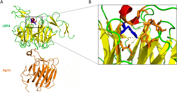
Figure 5.
3D view of β1 propeller domain of LRP4 in complex with agrin involving Tyr607 mutation. (A) Backbone representation extracted from the crystal structure of LRP4, in complex with agrin (colored in orange), using PDB accession number 3V64. Tyr607 mutation is colored in blue. (B) Zoom view of the side chain interactions between Tyr607 (colored in blue) and neighbor residues (colored in orange). Inter side-chain distances lower than 5 Å are indicated by dash lines. For A and B figures were extracted from PyMol software.

