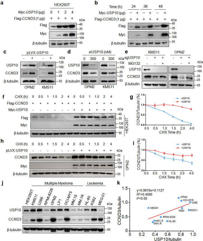
Fig. 3. USP10 stabilizes CCND3 in MM cells.
a A CCND3 plasmid was co-transfected with increased USP10 into HEK293T cells for 48 h, followed by IB assay. b CCND3 and USP10 were co-transfected into HEK293T cells for 24 to 48 h, followed by cell lysate preparation and IB assays. c OPM2 and KMS11 cells were infected with lentiviral USP10 for 72 h, followed by IB assays as indicated. d USP10 was knocked down from OPM2 and KMS11 cells by specific siRNA for 48 h, followed by IB assays. e USP10 was knocked out by its sgRNA, followed by MG132 treatment. The cell lysates were then subjected to IB assays. f USP10 and CCND3 were co-transfected into HEK293T cells for 36 h, followed by CHX treatment at indicated periods before collecting for IB assay. g The blots in f was subjected to desitometry for CCND3 expression against GAPDH. h The USP10 lentivirus was infected into OPM2 cells for 48 h, followed by CHX treatment at indicated periods before collecting for IB assay. i The blots in h was subjected to densitometry for CCND3 expression against GAPDH. j Cell lysates from cell lines were subjected to IB assays against CCND3 and USP10. k The relationship analysis between the CCND3 and USP10 expression levels from i.

