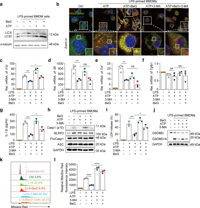
Fig. 4. BeG promotes mitophagy to inhibit NLRP3 inflammasome activation.
a Western blotting analysis of LC3B in BMDMs treated as indicated in the image. b Confocal microscopy of BMDMs pre-treated with BeG stimulated with LPS and treated with ATP, stained for LC3 with Mitotracker. Scale bars, 25 μm. c–f Effects of BeG, LPS, 3-methyladenine (3-MA, 5 mM), and ATP stimulation on level of Il1b, Il6, Tnf, Tgfb1 microRNA (mRNA) in BMDMs (n = 3 per group). g Measurement of IL-1β in BMDMs (n = 3 per group). h Immunoblot analysis of protein levels of core NLRP3 inflammasome components (NLRP3, ProCasp1, and ASC) and NLRP3 inflammasome activation-related proteins (Casp1-p10) from the BMDMs lysates and culture supernatants. i LDH release in culture supernatants (n = 3 per group). j Immunoblot analysis of GSDMD-N formation in BMDMs. k, l J774A.1 cells were pre-treated with BeG, treated with LPS with or without 3-MA (5 mM), stimulated by ATP, and then stained with MitoSOX and analyzed by flow cytometry (n = 3 per group). Data shown as mean ± SD (*P < 0.05, **P < 0.01, ***P < 0.001, ****P < 0.0001; ns not significant).

