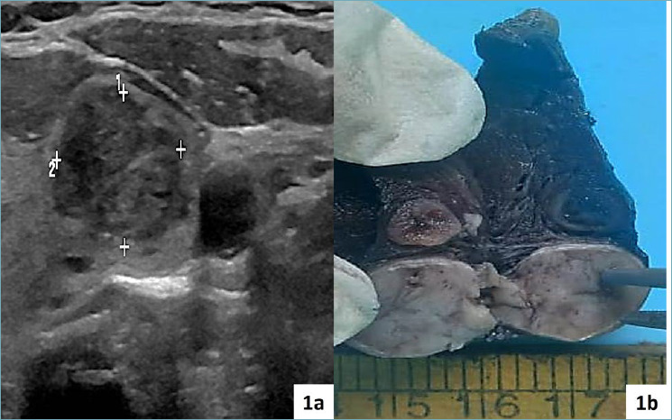
Figure 1.

(a) Ultrasonography of the left thyroid shows a solid nodule measuring approximately 1.5 cm with few areas of breakdown. Background thyroid showed few nodules of goitre. (b) The gross image of the left thyroid gland shows a well-defined capsulated solid whitish nodule with few areas of breakdown/cystic change. Background thyroid shows spongiform nodules of goitre.
