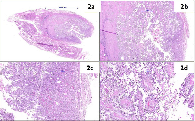
Figure 2.

(a) Scanner view of the thyroid nodule showing complete capsule of the nodule (HE40x); (b) Histology shows a well-encapsulated tumour composed of central papillary architecture and peripheral follicular architecture (HE100x); (c) interphase of the papillary and follicular architecture of the tumour (HE100x); (d) papillary adenocarcinoma morphology representing the lung tumour (HE200x).
