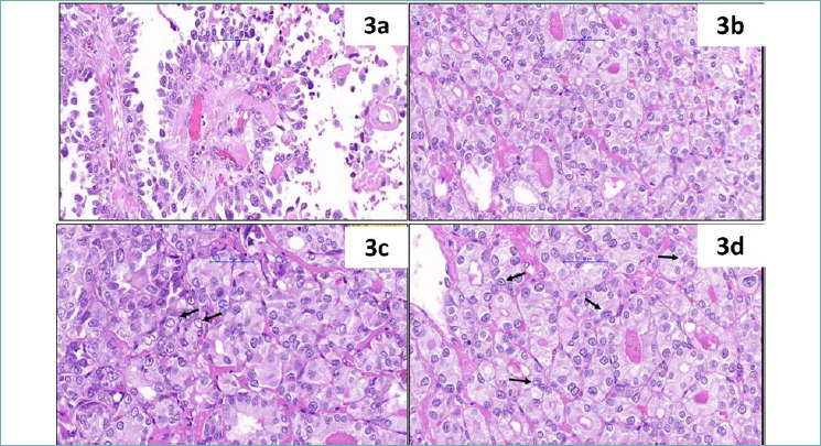
Figure 3.

(a) Well-formed papillae lined by tumour cells with high nuclear grade and moderate cytoplasm (HE400x); (b-d) follicular pattern of the tumour at the periphery showing mild nuclear enlargement and pale nuclei/ nuclear clearing seen in (c) (black arrow) along with nuclear grooves in (d) (black arrows) (HE400x).
