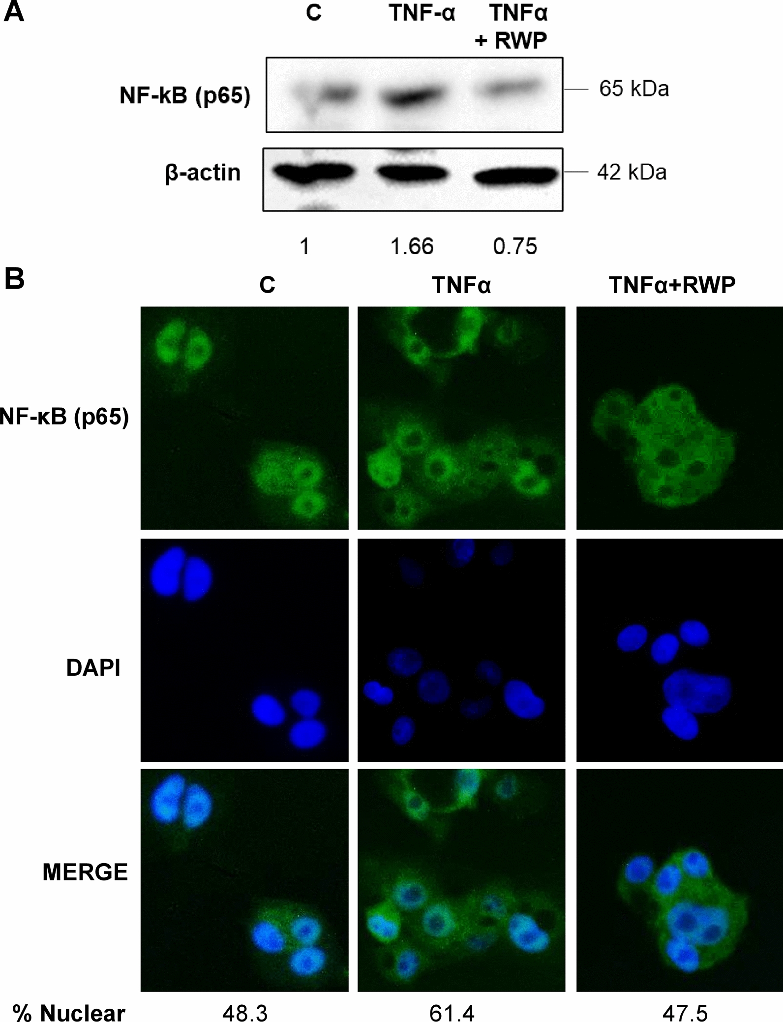
Fig. 5.

Effect of RWP on NF-kB in TNFα triggered hepatocytes. HH were triggered by 5 ng/mL TNFα in the absence (TNFα) or in the presence of 200 μg/mL RWP (TNFα + RWP). A Immunoblots were performed by using specific antibodies against subunit p65 of NF-κB and β-actin. Immunolabeled protein bands were normalized to β-actin by means. Values obtained are reported under western blot images. Protein expression levels in untreated HH (C) were taken as 1. B The cellular localization of subunit p65 of NF-kB was identified by immunocytochemistry experiments using a specific antibody. DAPI was used for nuclear staining. Photomicrographs typical of those taken three separate experiments are shown. In A and B, data are representative of at least 3 independent experiments. % Nuclear = [Total Nuclear Intensity/(Total Cytoplasmic Intensity + Total Nuclear Intensity)] × 100
