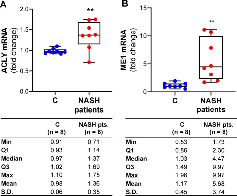
Fig. 7.
ACLY and ME1 mRNA expression levels in study population. Real time PCR experiments were performed to quantify ACLY (A) and ME1 (B) gene expression in macrophages from 8 healthy subjects (C) and 8 NASH patients. Data are shown as box plots, where the horizontal line within the boxes is the median, the boxes are the first and third quartiles, and the bars outside the boxes represent the minimum and maximum values. Dots indicate the fold changes of mRNA from each subject enrolled. Differences were significant according to Student’s t-test (**p < 0.01)

