Fig. 10.
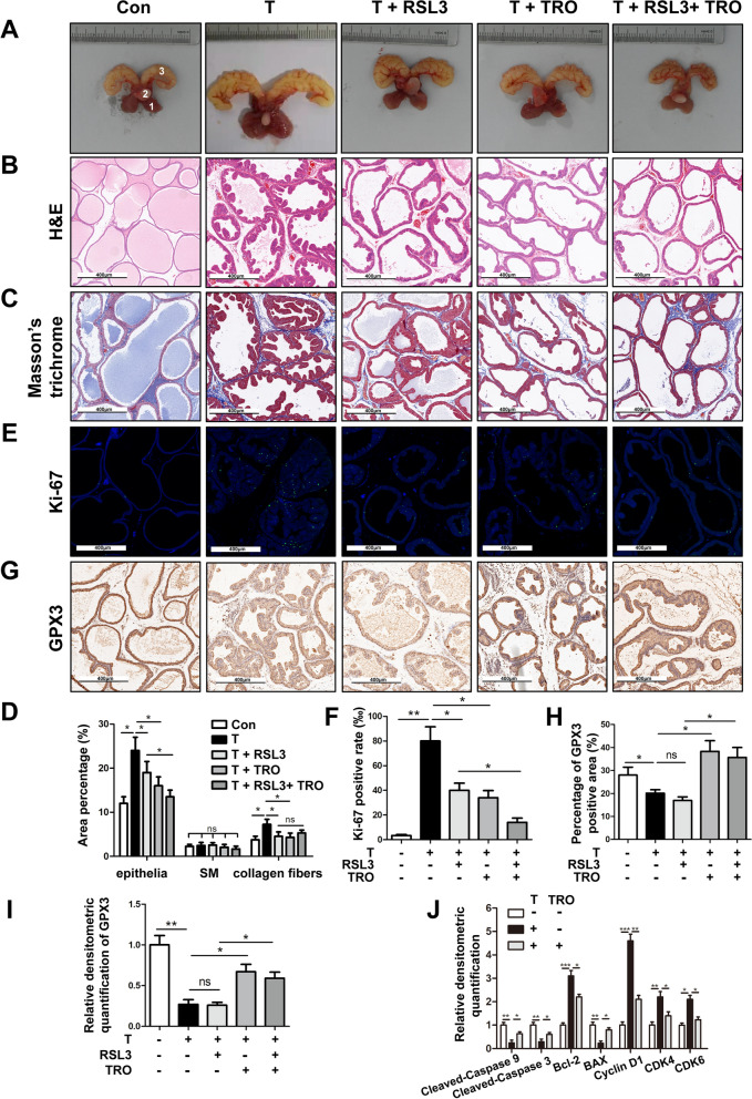
Effect of GPX3 on cell proliferation, cell cycle and apoptosis of prostate in vivo. A The rat urogenital tissues from Con, testosterone (T), T + RAS-selective lethal 3 (RSL3), T + troglitazone (TRO) and T + RSL3 + TRO-treated rats. (1) ventral prostate, (2) bladder and (3) seminal vesicle. B Representative H&E staining of rat prostates for each treatment group. Scale bars: 400 μm. C Masson’s trichrome staining of rat prostates for each treatment group; prostate epithelial cells were stained auburn, smooth muscle (SM) cells were stained red, and collagen fibers were stained blue. Scale bars: 400 μm. D Quantification of Masson’s trichrome staining. E The Ki-67 staining for the prostates from each group rats, respectively. DAPI (blue) and fluorescence-labeled images (green) are merged. Scale bars: 400 μm. F The bar graph for Ki-67-positive rate (%). The results showed the proliferative activity of prostate in T-BPH rats was significantly enhanced and could be inhibited by RSL3 or TRO. G Representative IHC images of GPX3 in each treatment group. Scale bars: 400 μm. H The quantitative analysis of GPX3 expression in each group rat prostates. I Relative densitometric quantification of GPX3 in prostate of each treatment group. The results indicated that TRO successfully promoted the expression of GPX3 in rat prostate. J Relative densitometric quantification of Cleaved-Caspase 9, Cleaved-Caspase 3, Bcl-2, BAX, Cyclin D1, CDK4 and CDK6 in prostate of Con, T, and T + TRO-treatment group, and the decrease of apoptosis and the acceleration of G0/G1 phase in the prostate of T-BPH rats could be inhibited by GPX3 upregulation in vivo. *p < 0.05, **p < 0.01, ***p < 0.001 and ns means no significant difference

