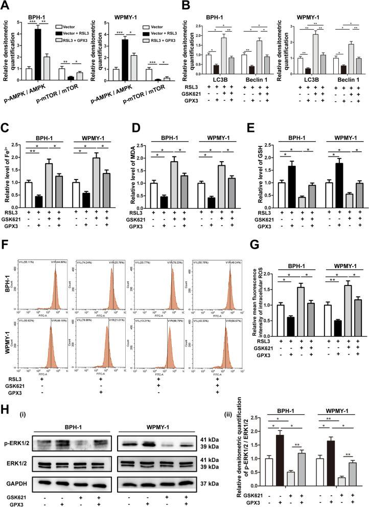
Fig. 8.
GPX3 antagonized autophagy-related ferroptosis and activated ERK1/2 with the involvement of AMPK A Relative densitometric quantification of proteins (AMPK, p-AMPK, mTOR and p-mTOR) in BPH-1 and WPMY-1 cells treated with RSL3 (0.5 μM) or GPX3 plasmid, and GPX3 overexpression reversed the up-regulation of p-AMPK and down-regulation of p-mTOR caused by RSL3 treatment. B Relative densitometric quantification of proteins (LC3B and Beclin1) in BPH-1 and WPMY-1 cells treated with GPX3 plasmid or GSK621 (20 μM), and GPX3 overexpression reversed the up-regulation of autophagy caused by GSK621 treatment. The content of iron (C), MDA (D) and GSH (E) in BPH-1 and WPMY-1 cells treated with GPX3 plasmid or GSK621 (20 μM) were detected by colorimetric assay kit. F DCFH-DA fluorescent probe was used to analyze the accumulation of ROS in BPH-1 and WPMY-1 cells treated with GPX3 plasmid or GSK621 (20 μM). G Statistical analysis of mean fluorescence intensity (MFI) of DCFH-DA. The results indicated that GPX3 overexpression reversed the up-regulation of autophagy-related ferroptosis caused by GSK621 treatment. H Immunoblot assay (i) and relative densitometric quantification (ii) of ERK1/2 and p-ERK1/2 in BPH-1 and WPMY-1 cells treated with 20 μM GSK621 or GPX3 plasmid showed that GPX3 regulated ERK1/2 signaling with the involvement of AMPK pathway. GAPDH is used as loading control. *p < 0.05, **p < 0.01, ***p < 0.001

