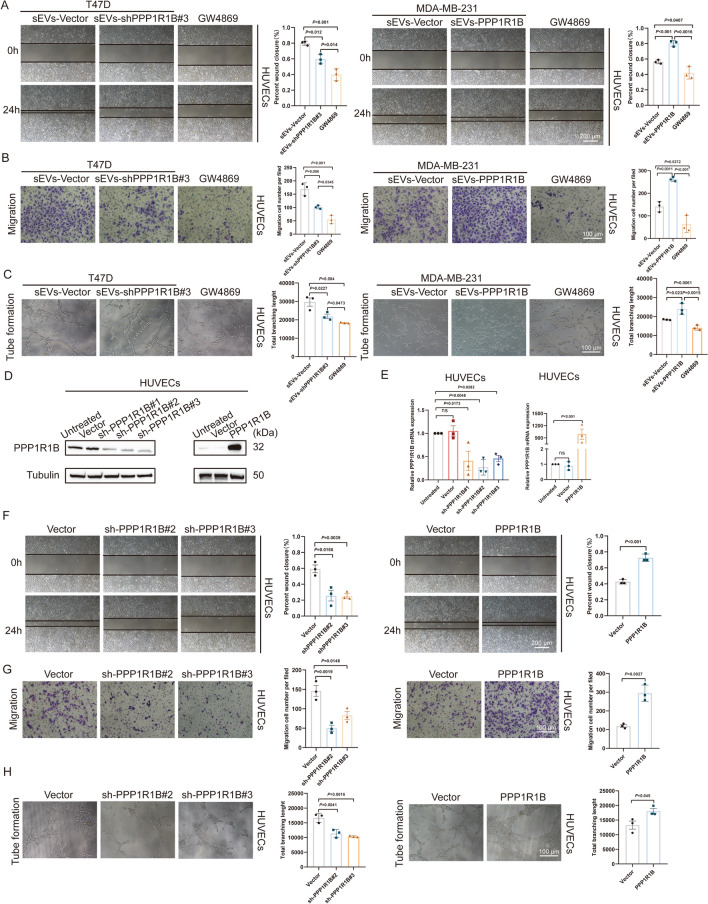
Fig. 7.
PPP1R1B promotes HUVECs angiogenesis. A, B The role of T47D and MDA-MB-231 cells derived sEVs cells on the migratory capacity of HUVECs. Representative micrographs of the wound healing and transwell assays. Scale bars, 200 µm and 100 µm. C Tube formation of HUVECs co-cultured with the sEVs-Vector, PPP1R1B-knockdown/overexpressed sEVs and GW4869. Scale bar, 100 µm. D Western blot analysis of PPP1R1B in HUVECs after transfection. E The expression of PPP1R1B was analyzed by qRT-PCR. F, G Wound healing and transwell assays were used to evaluat the migratory capacity of HUVECs after knockdown and overexpression of PPP1R1B. Scale bar, 200 µm and 100 µm. H Tube formation was performed to measure the angiogenic function of PPP1R1B in HUVECs. Scale bar, 100 µm. Data were the means ± SD of three experiments. The significant difference was evaluated with one-way ANOVA followed by the Bonferroni post hoc (A–C). Statistical significance was determined by a two-tailed unpaired t-test (F–H)

