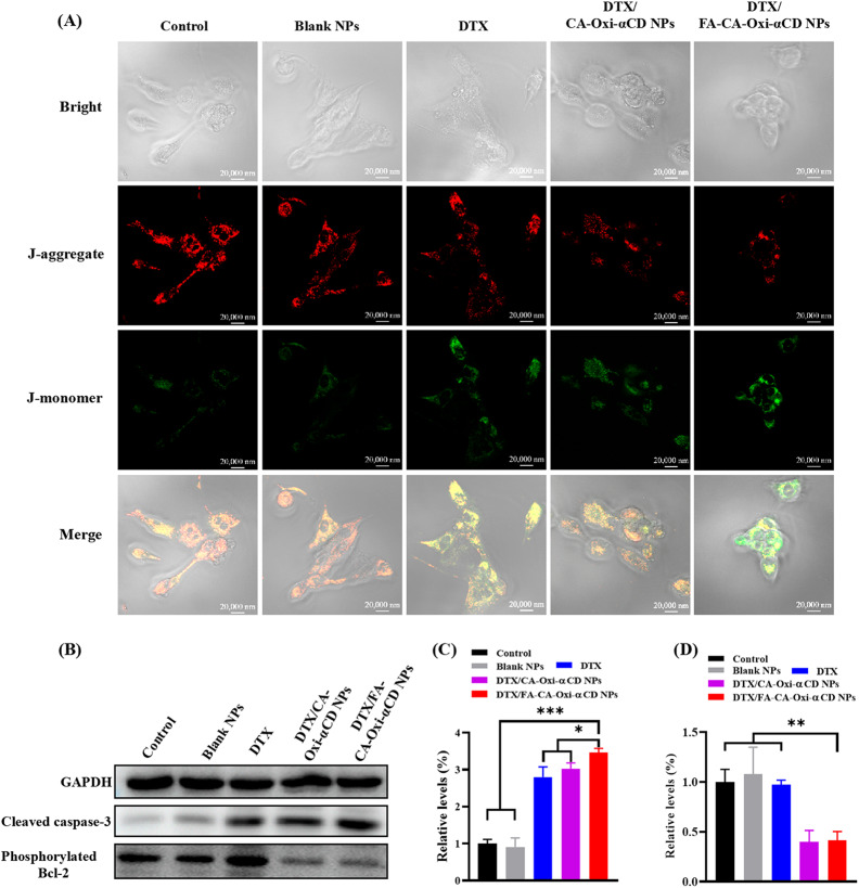
Fig. 4.
Mitochondrial damage-induced apoptosis of 4T1 cells by control, Blank NPs, DTX, DTX/CA-Oxi-αCD NPs and DTX/FA-CA-Oxi-αCD NPs, respectively. (A) CLSM images of JC-1 stained cells after different treatments. Scale bar represents 20 μm. (B) Western blot analysis for the expression of cleaved caspase-3 protein, phosphorylated Bcl-2 protein in 4T1 cells with different treatments for 48 h. (C) Quantitative analysis of cleaved caspase-3 protein expression in 4T1 cells. (D) Quantitative analysis of phosphorylated Bcl-2 protein expression in 4T1 cells. *p < 0.05, **p < 0.01, ***p < 0.001 compared with DTX/FA-CA-Oxi-αCD NPs (n = 3)

