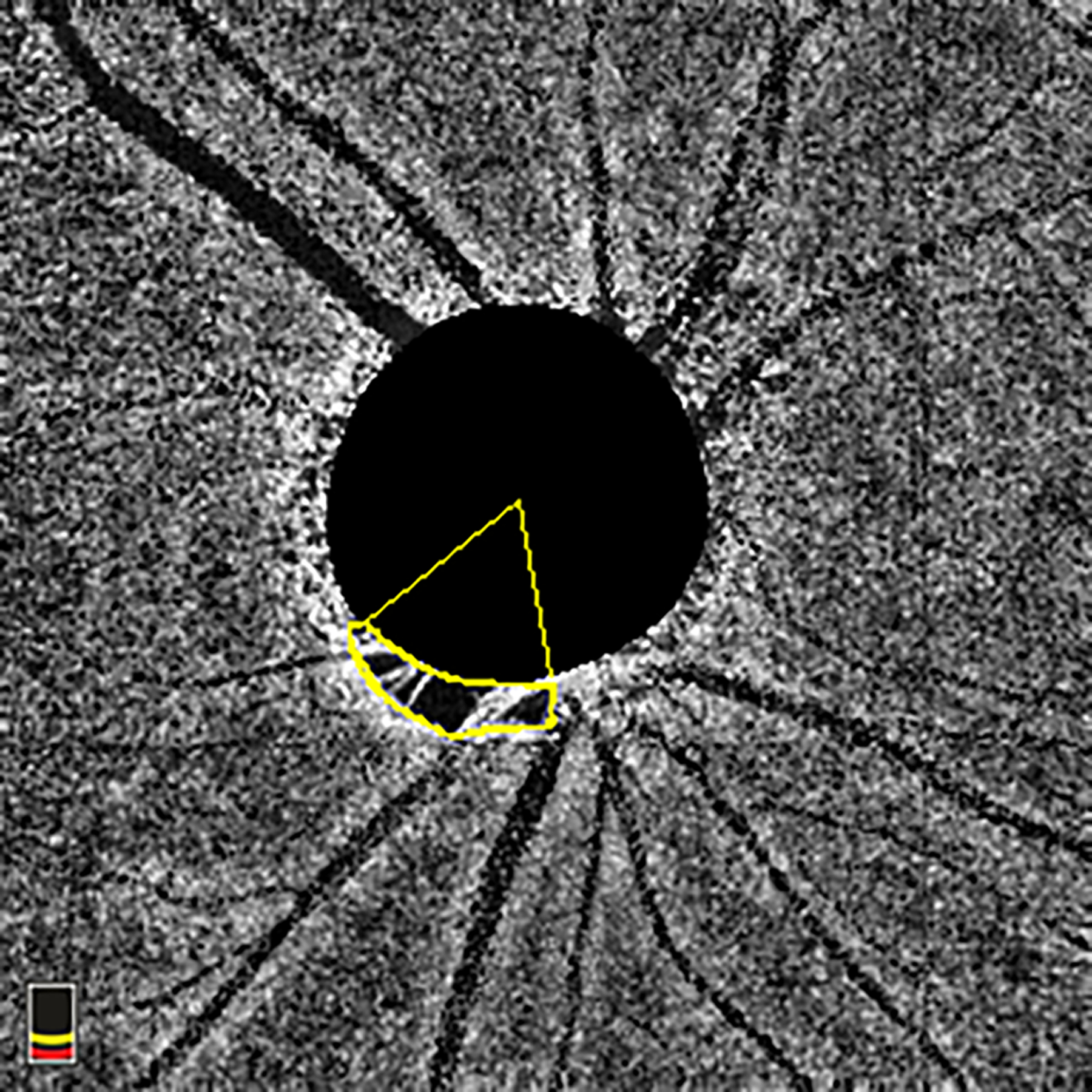
Figure 1.

En-face choroidal vessel density image showing Choroidal Microvasculature Dropout (MvD) area and angular circumference. MvD area was manually outlined using ImageJ software. The two points at which the extreme borders of MvD area met the optic nerve head (ONH) border were identified and defined as angular circumferential margins. The angular circumference was Then determined by drawing two lines connecting the ONH center to the angular circumference margins of the MvD. MvD = microvasculature dropout; ONH = optic nerve head.
