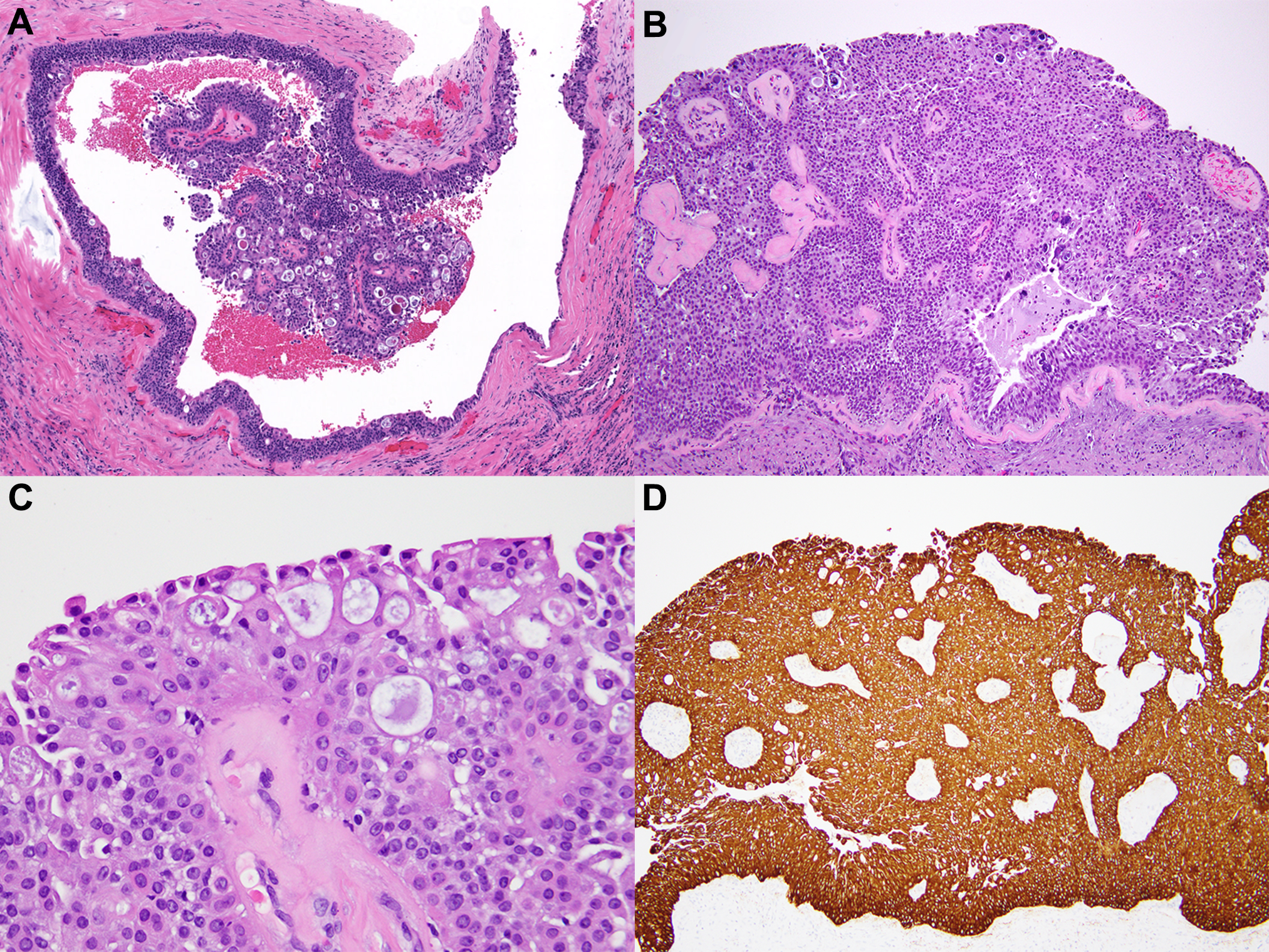
Figure 4.

Nodular cystic hidradenoma. Core biopsy (A), diagnosed outside as DCIS, was called atypical papillary lesion. Final diagnosis was rendered on the excision specimen (B) with consultation from a dermatopathologist. High power view shows a mixture of cell types (C). The cells are diffusely positive for high molecular weight cytokeratin (34βE12 shown in D).
