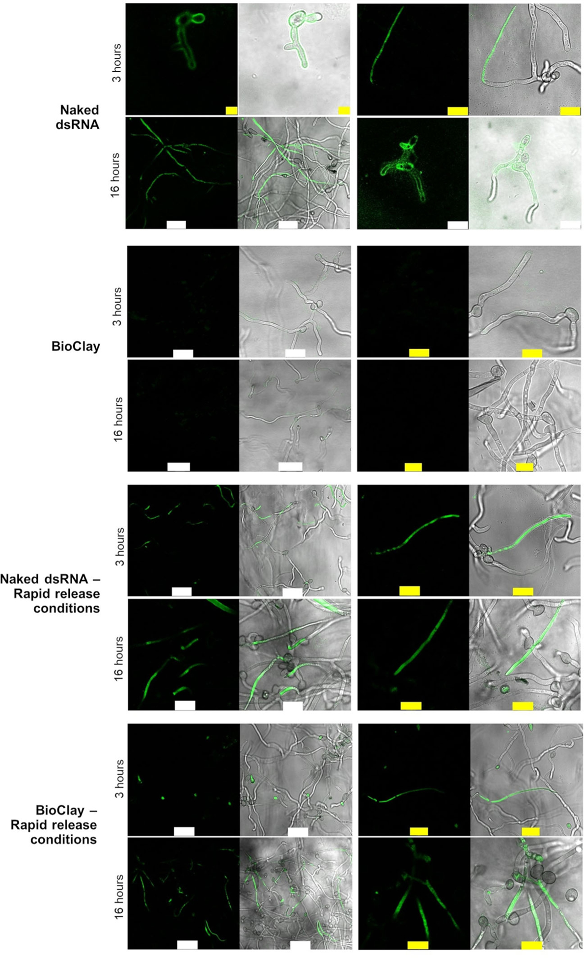
Figure 1. Fungal cells can take up dsRNA from BioClay™ after dsRNA release.

The images show the confocal laser scanning microscopy (CLSM) analysis of germinated spores 3 and 16 h after being treated with fluorescein-labeled dsRNA (YFP or BcDCL1/2). The top group of rows show that Botrytis cinerea can take up the labeled dsRNA on PDA medium at pH ~5.8 after 3 and 16 h. The second group of rows show that dsRNA cannot be taken up when the dsRNA is in the BioClay complex after 3 h. The third group of rows show the rapid release protocol does not affect dsRNA uptake when the dsRNA is in a naked form. The fourth group of rows show that dsRNA can be taken up under conditions that release the dsRNA from the BioClay complex. Micrococcal nuclease (MNase) was added 30 min before capturing the images to remove the external dsRNA, which was not taken up by fungal cells. However, because BioClay provides protection against nucleases, traces of fluorescent signals are observed at the periphery of the fungal cells. White bar = 25 μm, yellow bar = 10 μm.
