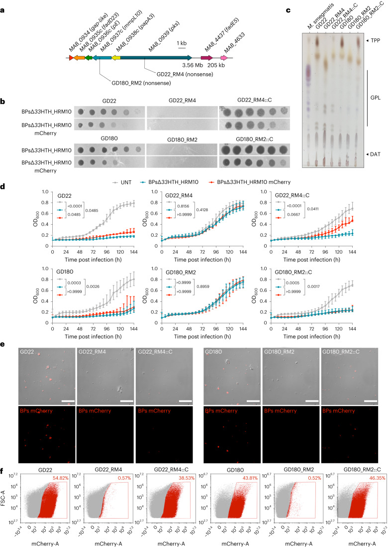
Fig. 4. TPPs are essential for BPs∆33HTH_HRM10 to lyse M. abscessus.
a, Representation of the M. abscessus TPP locus showing mutations affecting the clinical strains studied. b, Phages were spotted as tenfold serial dilutions onto clinical strains (GD22 and GD180), spontaneous resistant mutants (RM) and complemented strains (::C). Plates were incubated for 2–3 d at 37 °C before imaging. The assay was repeated at least three times and a representative experiment is shown. c, TLC analysis of total lipids extracted from M. abscessus clinical strains, resistant mutants and complemented strains. Eluent: CHCl3/CH3OH (90:10 v/v). Anthrone was sprayed on the plates to visualize the lipid profile, followed by charring. d, Liquid growth of the strains with BPs∆33HTH_HRM10 or BPs∆33HTH_HRM10 mCherry (MOI 10) or without phage (untreated; UNT) monitored every 6 h for 6 d at 37 °C in 7H9/OADC supplemented with 1 mM CaCl2. Data are plotted as the median ± interquartile range of three independent experiments done in triplicate. Statistical analysis conducted to compare the differences at 144 h between strains was done with a two-sided Dunn’s multiple comparisons test, with P values indicated. e, Representative fields of M. abscessus clinical strains infected with BPs∆33HTH_HRM10 mCherry (MOI 10) for 4 h at 37 °C before fixation. Infected bacilli appear in red. These results were obtained at least two times. Scale bars, 30 µm. f, Flow cytometry data represented as dot plot show the percentage of bacilli infected with the BPs∆33HTH_HRM10 mCherry fluorophage relative to the study population. This assay was conducted at least twice.

