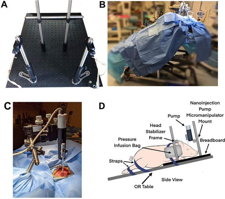
Figure 1.
Surgery for cortical AAV injection and installation of the cranial imaging window. (A) The pig’s head was secured in the breadboard with two vertical bars next to their snout and the horizontal bar pressing on the dorsal portion of its snout. Pressure infusion bags were inflated between the pig’s head and the back two vertical bars with the area between the chest and front legs pressed into the vertical bars. The pig’s front legs were tied to the board and the board tied to the OR table. Once the pig was draped, a sterile bar was placed horizontally between the back two vertical bars. (B) Photograph of the pig in position for AAV injection and imaging port implant. The pig was placed at a 30–45° angle to encourage the brain to fall away from the dura for implantation of PDMS avoiding cortical damage. (C) The sterile cross bar added to the vertical bars after draping the animal. Then, the sterile injector and manipulator were mounted to the cross bar. (D) Schematic depiction of pig positioning shown in (B).

