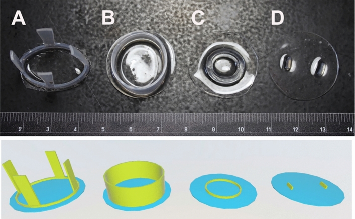
Figure 2.
The evolution of the dural substitute (and corresponding 3D renderings). (A) PDMS with silicone straps worked well but prevented later cortical impact. (B) PDMS with “top hat” portion to fill the edges of the burr hole. This version was difficult to place without causing cortical trauma. (C) PDMS with flat silicone ring. The ring didn’t provide much advantage and limited the area for 2P imaging. (D) Flat PDMS with ridges to identify the center of the PDMS. This iteration was relatively easy to install, allowed centering of the PDMS, and could withstand injections and cortical impact.

