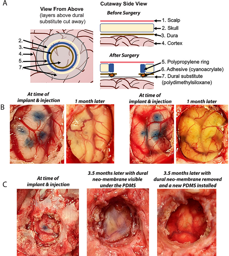
Figure 3.
Cranial imaging window. (A) The cranial imaging window system from the top and the side that prevented the adhesion of the healing of the dura onto the cortex that obfuscated the surface of the cortex as well as tissue growth from the burr hole of the skull. (B) Two burr holes displaying the PDMS overlaying the cortex immediately after AAV injection and the pristine window 1 month later. (C) The cranial window at installment, after 3.5 months with dural neomembrane obscuring the cortex, and immediately after dural neo-membrane removal and PDMS re-installation. This iteration of the cranial window had an adjacent subdural electrode strip that entered posterior to the imaging window.

