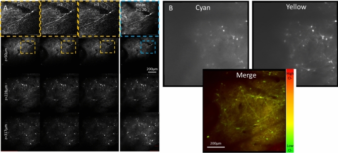
Figure 6.
Imaging in vivo intraneuronal chloride of the cortex. (A) The heartbeat triggered acquisition of a fast z-stack spanning 600 × 600 × 400 µm provided stable images of the fluorescent protein-transduced neurons in vivo. Because these volumes were acquired between heartbeats, individual iterations (first 3 columns) could be averaged together to increase dynamic range and decrease shot noise in the final image stacks (4th column). (B) Merging the cyan and yellow fluorescent protein signals from SuperClomeleon provides a pseudo-colored imaged of intracellular chloride.

