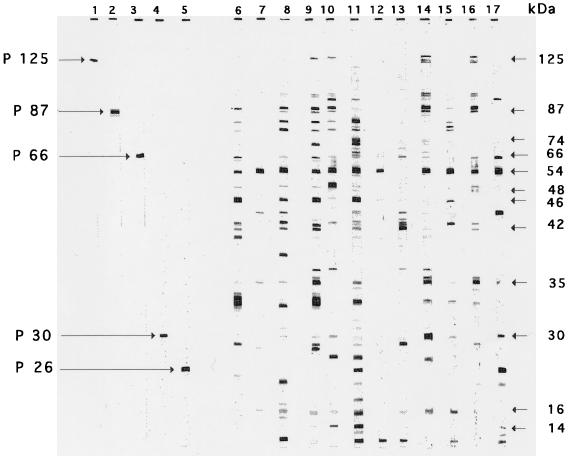
FIG. 1.
Immunoblot patterns obtained with a saline extract from H. pylori ATCC 43579 with five rabbit sera raised against purified antigens from H. pylori (lanes 1 to 5) and with 12 selected sera from patients infected with H. pylori (lanes 6 to 17). Molecular masses are indicated on the right. The arrows indicate immunoreactive bands corresponding to the antigens p125, p87, p66, p30, and p26. The figure shows a scan of the original nitrocellulose strips.

